This post was written based on the appearance of Dr. Robert Bednarek, Micrographic Surgery and Dermatologic Oncology, Parkview Packnett Family Cancer Institute, on the program, PBS HealthLine.
Overview
Melanoma, like any cancer, is characterized by abnormal cell growth. In this case, the cells come from the melanocytes.
The skin is made up of three layers: the epidermis, the dermis, and the hypodermis (fat). Normally, melanocytes reside in the bottom portion of the top layer of skin (epidermis), and they play a really important role in protecting the skin. Most people associate melanocytes with tanning. The increased pigment that comes with tanning is melanocytes reacting to ultraviolet radiation, and the body attempting to protect the rest of the skin. Melanoma occurs when the skin cells mutate or grow abnormally.
What increases risk?
Risk factors for melanoma can be divided into two groups: genetic factors, those that can't be altered, and environmental factors, which can be controlled or modified.
When discussing environmental factors, ultraviolet radiation, whether from the sun or tanning beds, is the tried-and-true number one most controllable factor for skin cancer in general, and for melanoma in particular.
Factors that cannot be controlled, such as having blue eyes, fair skin and a tendency to freckle, can also increase vulnerability to UV exposure. Additionally, those who are born with a decreased immune system or have developed immunosuppression due to a chronic illness may also face a higher risk of melanoma.
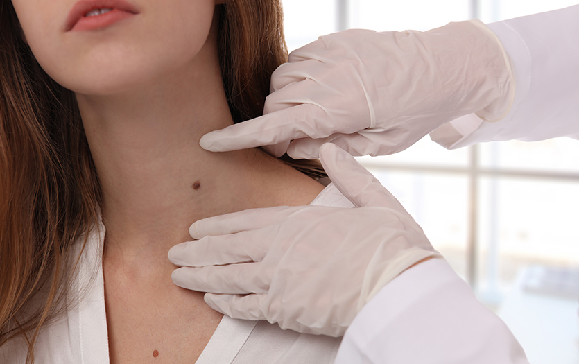
How is melanoma diagnosed?
Usually melanoma develops on the skin, but it can also appear in other areas of the body that receive little sun exposure, including:
-
Eyes
-
Nail beds of the fingers and toes
-
Mucous membranes in the nose, sinuses, mouth, digestive tract and urinary tract
When a provider is evaluating a spot or a mole of concern, they reference the A-B-C-D-E checklist:
-
A for Asymmetry. If cut in half, would the mole look the same on both sides?
-
B for Border. Does the mole have a clear-cut border? Is it evident where the border starts and stops? In melanoma, it will have an irregular or more scalloped appearance.
-
C for Color. In general, moles appear in one or two uniform colors; however, melanoma often exhibits three, four, or even five different colors. Typically, normal moles are white or slightly off-white, tan or brown. In contrast, melanoma can present with dark browns, light browns, reds, blues and whites. It doesn't adhere to a specific color range.
-
D for Diameter. Anything larger than 6 mm, which equates to about the size of a pencil eraser, may need further evaluation.
-
E for Evolution. When evaluating moles, especially in individuals over 40, a change in a mole raises concerns about malignancy.
The mainstay of diagnosing melanoma is performing a biopsy. Samples are typically obtained under general anesthesia, and when possible, the provider will remove the entire area of concern. This allows the dermatopathologist the best opportunity to make an accurate diagnosis and assess the depth of the melanoma. A patient's prognosis and the subsequent stage are most closely correlated with melanoma thickness. The thinner the melanoma, the better the prognosis.
Like other cancers, melanoma can metastasize or spread to different areas of the body. Depending on the depth measured on pathology, it may be recommended to have further testing such as a sentinel lymph node biopsy, as that is likely the first place it will spread beyond the skin.
It's also important to refrain from scratching or picking at moles or skin growths. In melanoma staging, ulceration or breakdown of the skin's outer layer is an indicator of tumor behavior. Self-inflicted damage may cause inaccurate staging results.
Treatment
Staging often dictates the course of disease management. Early detection and diagnosis offer the best treatment options and outcomes. For very thin melanomas, the five-year survival rate is close to 99-100%. Patients can often receive treatment for these via office-based surgery. Surgical removal of melanoma, when that is an option, is the most effective treatment for localized early-stage tumors.
If the cancer has spread beyond the skin, a more aggressive treatment approach is necessary. This can include radiation, chemotherapy and immunotherapy in addition to surgical removal.
Prevention
Sun protection is one of the most effective methods for preventing melanoma and for individuals caring for themselves during and after treatment. The American Academy of Dermatology recommends using a water-resistant sunscreen with an SPF of 30 or higher. The sunscreen must be broad-spectrum, meaning it protects against both UVA (ultraviolet A) and UVB (ultraviolet B) rays. The difference in protection between SPF 30 and SPF 100 is only a few percentage points, despite the significant numerical increase. Applying sunscreen once a day is not enough when spending time in direct sunlight. Thoroughly reapply sunscreen every two hours or every 80 minutes if sweating or in the pool.
Learn more about our dermato-oncology team here.

.jpg)